In this post, we present a case report from Dr. Stephen Tuohy (Beacon Hospital Dublin, Ireland) of a patient suffering from ventricular tachycardia (VT). The magnetic resonance images (MRI) were analysed with ADAS 3D software to extract detailed information from the patient.
What are the patient characteristics?
The patient was a 61-year-old female with nonischaemic cardiomyopahty (NICM), complete AV block and recurrent VT. The patient had an implantable cardioverter-defibrillator (ICD) placed after the cardiac magnetic resonance acquisition. Moreover, she had a family history of VT and cardiomyopathy.
The VT morphology suggested an anterior RV site of origin. Previously, electrophysiologists attempted a VT ablation but was unsuccessful —weak match of pace-mapped to RVOT and empiric ablation unsuccessful—. The VT was suppressed with a drug approach based on amiodarone but some side effects appeared. Therefore, the medical team planned a redo for the VT ablation. An imaging guidance was requested to help with this challenging case.
Analysis with ADAS 3D software
Before the procedure, the DICOM images were imported into ADAS 3D software to be post-processed. The software generated a 3D model of the left ventricle and represented the adjacent anatomy.
Tissue is displayed in blue for non-enhanced tissue (healthy), in red for highly-enhanced tissue (core), and from light blue to orange represents the grey-area tissue. The white tube are the centerlines of the automatically detected border-zone corridors.

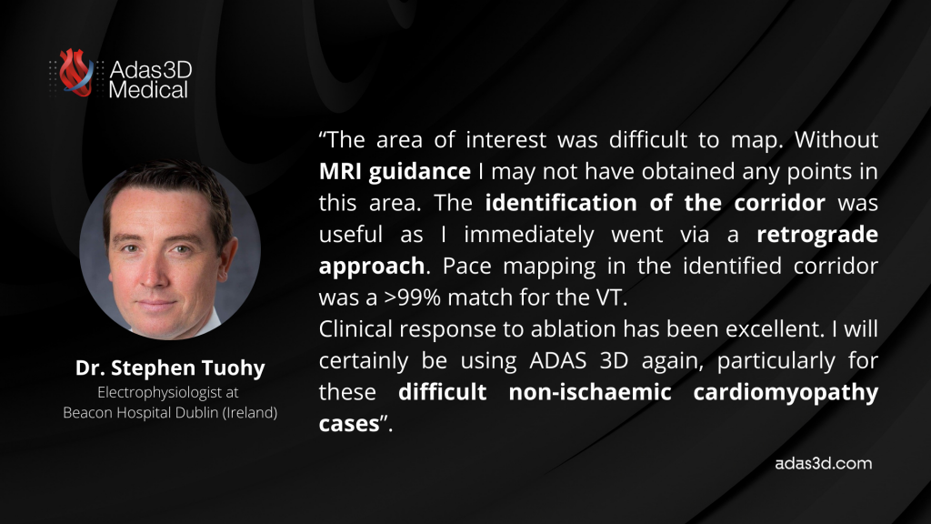
The location of the corridor helped to decide the retrograde approach for the procedure. Redo ablation was successful and the clinical response from the patient was excellent.
What does the expert say?

Thank you Dr. Tuohy for sharing this interesting case with us.
If you want to share your successful cases analysed with ADAS 3D, please fill in this form.
