Featured Cases
Cases
Scientific publications
Cases
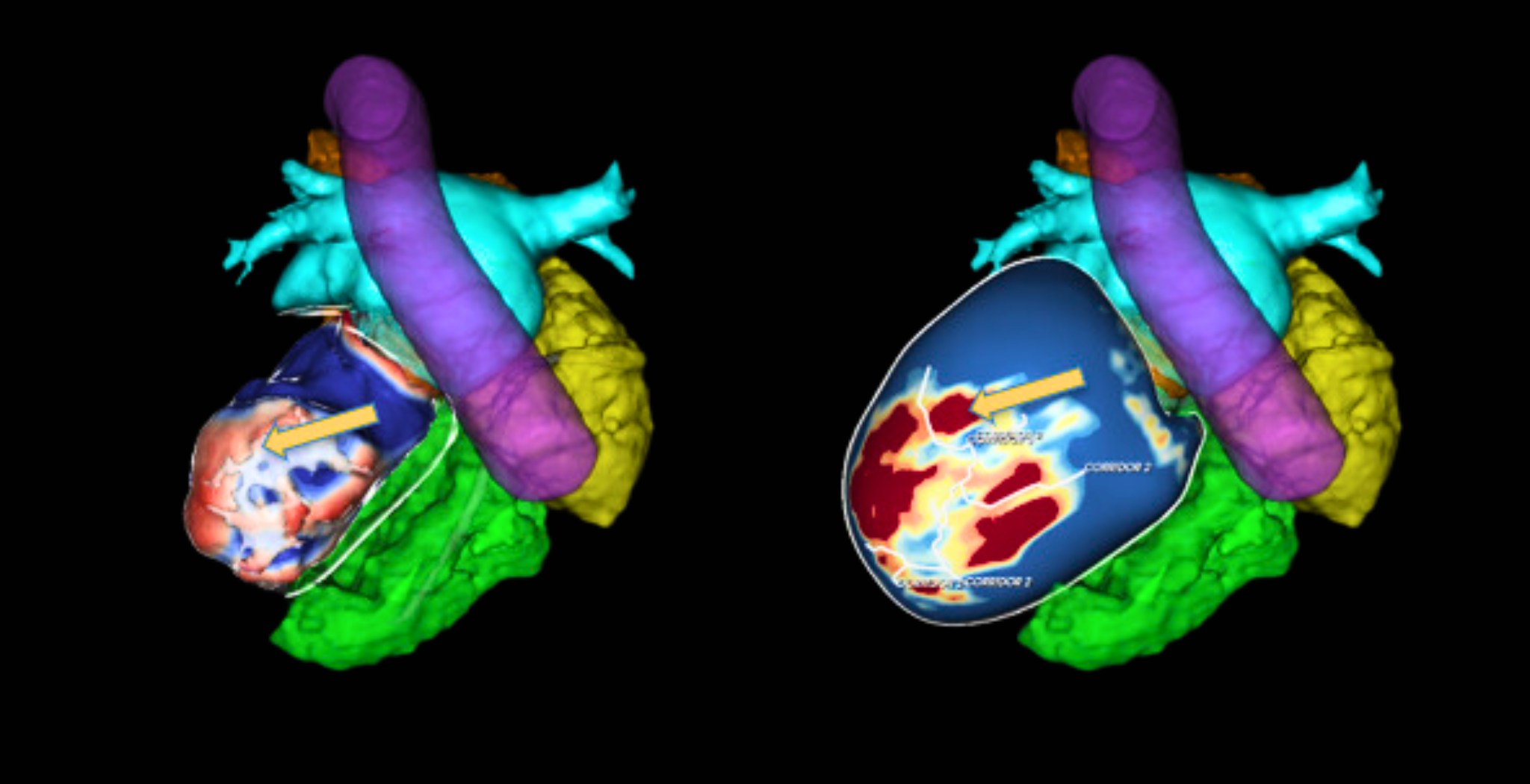
Case of VT Ablation in a patient with Mitral Valve Prosthesis using ADAS 3D model to plan the procedure.
We present a case from Dr. Alba Santos (Hospital Vall d’Hebron, Spain) of a patient suffering from ventricular tachycardia (VT). The magnetic resonance images (MRI) and computed tomography (CT) were analyzed with ADAS 3D software to extract detailed information about the patient. Description of the case We present a 50-year-old woman with a history of inferior myocardial infarction with no significant lesions…
Challenging case of recurrent Ventricular Tachycardia
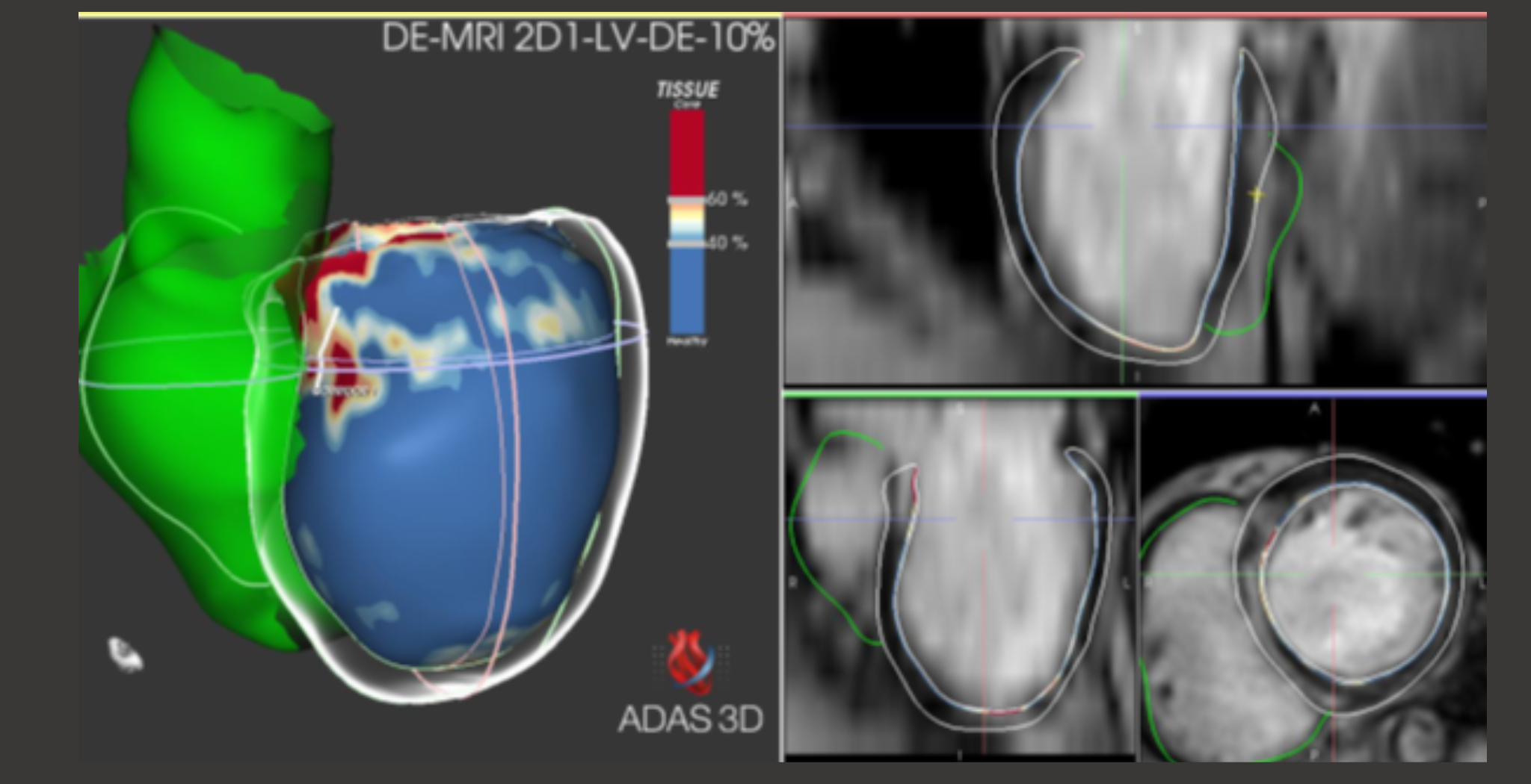
In this post, we present a case report from Dr. Stephen Tuohy (Beacon Hospital Dublin, Ireland) of a patient suffering from ventricular tachycardia (VT). The magnetic resonance images (MRI) were analysed with ADAS 3D software to extract detailed information from the patient. What are the patient characteristics? The patient was a 61-year-old female with nonischaemic cardiomyopahty (NICM), complete AV block and…
Case of VT procedure using ADAS3D with Ensite and HD-grid
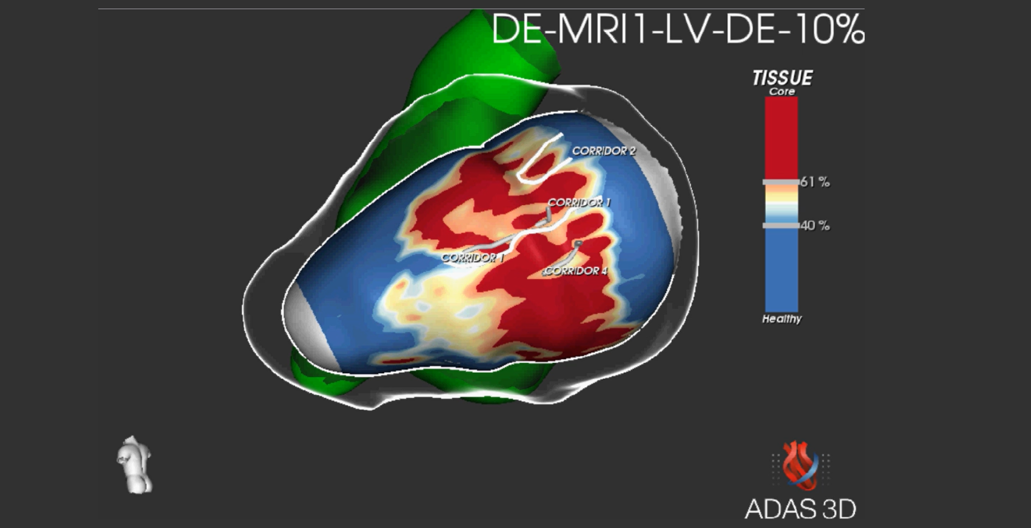
We present a case report from Dr. Javier Moreno (Hospital Ramón y Cajal, Spain) of a patient suffering from ventricular tachycardia. The MRI shows a scar in basal and middle segments of the inferior-lateral face of the left ventricle. Before the procedure The analysis of the DICOM was performed by ADAS 3D and the result showed an endocardial-transmural scar in…
Scientific publications
The following publications have used an ADAS 3D investigational version (not available for clinical use in the US) with not certified features. Interpretation of results should be done with caution as a scientific reference.
Left ventricle
Ablation of ventricular tachycardia using state-of-the art pre-procedural imaging, magnetic-based three-dimensional mapping and ultra-low temperature cryoablation technology
Liebregts M, Van Dijk VF, Boersma LVA, Balt JC. Heart Rhythm Case Reports 2022 https://doi.org/10.1016/j.hrcr.2022.02.013
Cardiovascular magnetic resonance determinants of ventricular arrhythmic events after myocardial infarction
Jáuregui B, Soto-Iglesias D, Penela D, Acosta J, Fernández-Armenta J, Linhart M, Ordóñez A, San Antonio R, Terés C, Chauca A, Carreño JM, Scherer C, Falasconi G, Prat-González S, Perea RJ, Mont L, Bosch X, Ortiz-Pérez JT, Berruezo A. Europace 2021; euab275 https://doi.org/10.1093/europace/euab275
Cardiac magnetic resonance to predict recurrences after ventricular tachycardia ablation: septal involvement, transmural channels, and left ventricular mass
Quinto L, Sanchez P, Alarcón F, Garre P, Zaraket F, Prat-Gonzalez S, Ortiz-Perez JT, Jesús Perea R, Guasch E, Tolosana JM, San Antonio R, Arbelo E, Sitges M, Brugada J, Berruezo A, Mont L, Roca-Luque I. Europace 2021;00:1-9 https://doi.org/10.1093/europace/euab127
Follow-Up After Myocardial Infarction to Explore the Stability of Arrhythmogenic Substrate: The Footprint Study
Jáuregui B, Soto-Iglesias D, Penela D, Acosta J, Fernández-Armenta J, Linhart M, Terés C, Syrovnev V, Zaraket F, Hervàs V, Prat-González S, Perea RJ, Morales-Ruiz M, Jiménez W, Lasalvia L, Bosch X, Ortiz-Pérez JT, Berruezo A. Journal of American College of Cardiology: Clinical electrophysiology 2020;6(2):207-18 https://doi.org/10.1016/j.jacep.2019.10.002
Ventricular scar channel entrances identified by new wideband cardiac magnetic resonance sequence to guide ventricular tachycardia ablation in patients with cardiac defibrillators
Roca-Luque I, Van Breukelen A, Alarcon F, Garre P, Tolosana J-M, Borras R, Sanchez P, Zaraket F, Doltra A, Ortiz-Perez J-T, Prat-Gonzalez S, Perea R-J, Guasch E, Arbelo E, Berruezo A, Sitges M, Brugada J, Mont L. Europace 2020;0:1-9 https://doi.org/10.1093/europace/euaa021
Cardiac Magnetic Resonance – Guided Ventricular Tachycardia Substrate Ablation
Soto-Iglesias D, Penela D, Jáuregui B, Acosta J, Fernández-Armenta J, Linhart M, Zucchelli G, Syrovnev V, Zaraket F, Terés C, Perea R-J, Prat-González C, Doltra A, Ortiz-Pérez J-T, Bosch X, Camara O, Berruezo A. JACC: Clinical electrophysiology 2020;6(4):436-447 https://doi.org/10.1016/j.jacep.2019.11.004
Left atrium
Late gadolinium enhancement-MRI determines definite lesion formation most accurately at 3 months post ablation compared to later time points
Althoff TF, Garre P, Caixal G, Perea R, Prat S, Tolosana JM, Guasch E, Roca-Luque I, Arbelo E, Sitges M, Brugada J, Mont L. Pacing Clinical Electrophysiology 2021;1-11 https://doi.org/10.1111/pace.14415
Personalized paroxysmal atrial fibrillation ablation by tailoring ablation index to the left atrial wall thickness: the ‘Ablate by-LAW’ single-centre study—a pilot study
Teres C, Soto-Iglesias D, Penela D, Jáuregui B, Ordónez A, Chauca A, Carreño JM, Scherer X, San Antonio R, Huguet M, Roque A, Ramírez C, Oller G, Jornet A, Palet J, Santana D, Panaro A, Maldonado G, De Leon G, Jiménez G, Evangelista A, Carballo J, Ortíz-Pérez JT, Berruezo A. EP Europace 2021; euab216 https://doi.org/10.1093/europace/euab216
Accelerated 3D Left Atrial Late Gadolinium Enhancement in Patients with Atrial Fibrillation at 1.5 T: Technical Development
Gunasekaran S, Haji-Valizadeh H, Lee DC, Avery RJ, Wilson BD, Ibrahim M, Markl M, Passman RS, Kholmovski EG, Kim D. Radiology: Cardiothoracic Imaging 2020;2(5):e200134 https://doi.org/10.1148/ryct.2020200134
Verification of threshold for image intensity ratio analyses of late gadolinium enhancement magnetic resonance imaging of left atrial fibrosis in 1.5T scans
Bertelsen L, Alarcón F, Andreasen L, Benito E, Salling Olesen M, Vejlstrup N, Mont L, Hastrup Svendsen J.International Journal of Cardiovascular Imaging 2020;36:513-520 https://doi.org/10.1007/s10554-019-01728-0
Left atrial fibrosis quantification by late gadolinium-enhanced magnetic resonance: a new method to standardize the thresholds for reproducibility
Benito E-M, Carlosena-Remirez A, Guasch E, Prat-González S, Perea RJ, Figueras R, Borràs R, Andreu D, Arbelo E, Tolosana J-M, Bisbal F, Brugada J, Berruezo A, Mont L. Europace 2017;19(8):1272-1279 https://doi.org/10.1093/europace/euw219
Simplified mapping and ablation of a scar-related atrial tachycardia using magnetic resonance imaging tissue characterization
Bisbal F, Andreu D, Berruezo A. Europace 2015;17(2):186 https://doi.org/10.1093/europace/euu307
ADAS 3D around the world
ADAS 3D around the world:
Subscribe to our newsletter
Discover the latest news and upcoming events related to ADAS 3D


